
Introduction: Understanding Knee Osteoarthritis and Its Diagnostic Challenges
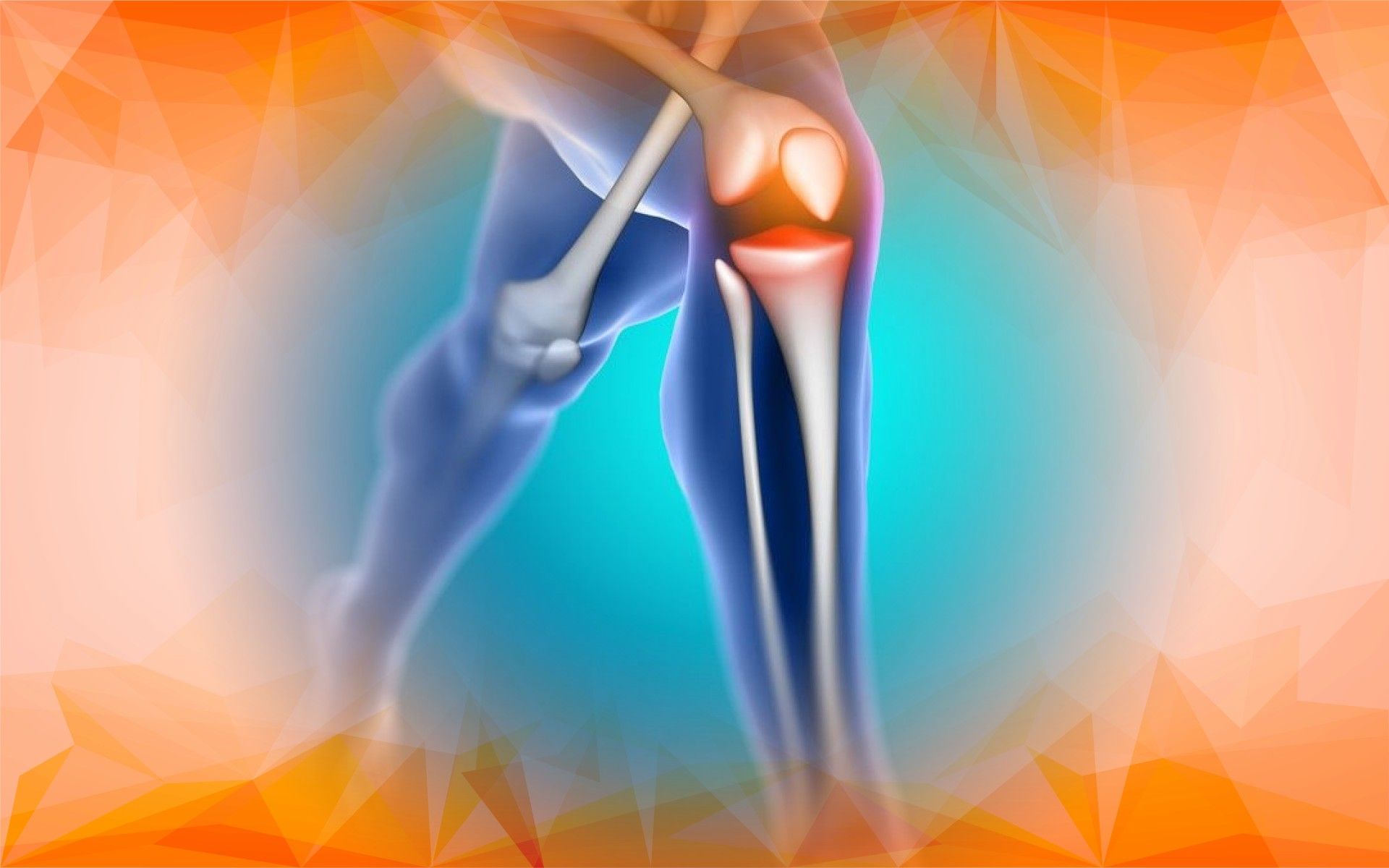
Knee osteoarthritis is a widespread condition where the cartilage cushioning the knee joint gradually breaks down. This often results in pain, stiffness, and difficulty moving—affecting millions of people globally. Early and accurate diagnosis is key for managing symptoms and slowing the progression of the disease.
Traditionally, knee osteoarthritis is diagnosed through a combination of physical exams and imaging tests, mostly X-rays. While helpful, these methods sometimes fail to catch subtle early changes and can have limitations. As Lane and Wallace (2002) point out, diagnosing osteoarthritis can be a process of elimination, since there aren’t standard blood or urine tests. In some cases, patients may even self-diagnose after comparing their symptoms (Machin, 2015). In this article, we’ll explore how innovative computer-based tools—specifically, certainty factor models—are working alongside traditional imaging techniques to make knee osteoarthritis diagnosis more accurate and timely.
Traditional Diagnosis: X-rays and the Kellgren-Lawrence Scale
When doctors suspect knee osteoarthritis , they start with the patient’s medical history, conduct a physical exam, and often order an X-ray. The most widely used method for interpreting these X-rays is the Kellgren-Lawrence (K-L) scale. This scale grades the severity of osteoarthritis based on signs like joint space narrowing and the formation of bone spurs.
Despite being the gold standard for decades, the K-L scale isn’t perfect. It’s sometimes difficult to distinguish between the subtleties of joint changes just by looking at a two-dimensional X-ray. Since X-rays can’t show every detail—especially in the early stages—crucial information may be missed. On top of that, osteoarthritis often coexists with other joint conditions, making the diagnostic process more complex (Lane & Wallace, 2002). Symptoms like persistent, dull pain that worsens with movement and eases with rest can overlap with other disorders too (Machin, 2015), adding another layer of challenge for doctors.
Why Traditional Methods Sometimes Fall Short
Relying solely on X-rays and the K-L scale can sometimes mean that minor but important changes inside the knee go unnoticed. Interpreting these images can be subjective, leading to missed or delayed diagnoses and less individualized care.
Recognizing these gaps, researchers have developed new tools to assist doctors in making more confident and precise diagnoses. These support systems are designed to provide a broader and clearer understanding of what’s happening inside the knee joint .
Introducing Certainty Factor Models: Smarter Diagnostic Tools
Certainty factor models represent a new wave of diagnostic support systems. Imagine them as smart digital assistants for doctors. These tools analyze a mix of information—such as patient symptoms, medical history, and imaging results—and calculate a “certainty score” that reflects how confident the system is in diagnosing osteoarthritis .
Instead of simply replacing the expertise of a doctor, certainty factor models augment it. By weighing all available evidence, these models help doctors make more accurate, consistent, and timely decisions while reducing guesswork.
Combining Traditional Imaging with Modern Technology
The most promising approach is to blend tried-and-true methods with these intelligent systems. While the Kellgren-Lawrence scale helps grade osteoarthritis severity, certainty factor models bring in more precision by considering a broader range of factors and offering a confidence level for the diagnosis.
Advanced imaging such as magnetic resonance imaging (MRI) adds even more detail. MRIs can reveal the state of cartilage , bone, and soft tissue that X-rays might miss. When certainty factor models analyze MRI data along with other patient information, doctors get a more complete—and often earlier—picture of knee health . For example, research shows that “feature learning from T2 maps has potential in uncovering information that can potentially better diagnose OA than simple averages or linear patterns decomposition” (Pedoia et al., 2019).
What Research Tells Us: Improved Accuracy and Patient Care
Recent studies are striking: diagnostic accuracy can exceed 90% when certainty factor models are used alongside traditional imaging and clinical information. These systems can sift through complex patterns in imaging and patient data to more accurately differentiate between the earliest signs of osteoarthritis and more advanced cases.
For instance, research using deep learning on MRI T2 data achieved impressive results: “DenseNet trained on raw T2 data obtained AUC = 83.44%, Sensitivity = 76.99%, Specificity = 77.94%”—outperforming older methods (Pedoia et al., 2019).
This leap in diagnostic power means doctors can better tailor treatments to a patient’s specific needs, helping prevent symptoms from worsening and allowing people to maintain their lifestyles for longer. Early and precise diagnosis is crucial, as untreated osteoarthritis can eventually lead to significant mobility issues and lifestyle limitations (Machin, 2015).
Looking Ahead: The Future of Osteoarthritis Diagnosis
In summary, while X-rays and grading systems like Kellgren-Lawrence remain valuable, they’re no longer enough on their own. Certainty factor models and expert systems introduce objective, data-driven analysis, providing extra confidence and clarity.
Blending these modern tools with time-tested methods allows healthcare providers to diagnose knee osteoarthritis earlier and more reliably. This integrated, smarter approach means better outcomes for patients—helping them stay active, maintain independence, and enjoy a higher quality of life.
The future of knee osteoarthritis diagnosis is here: combining expert insight with intelligent technology to offer the best care possible.
References
Lane, N. E., & Wallace, D. J. (2002). How Is Osteoarthritis Diagnosed? In (pp. 47-52). Oxford University Press.
Machin, A. (2015). A self-diagnosed case of osteoarthritis. InnovAiT: Education and inspiration for general practice, 9(9), 571-572. https://doi.org/10.1177/1755738015602276
Pedoia, V., Lee, J., Norman, B., Link, T. M., & Majumdar, S. (2019). Diagnosing osteoarthritis from T2 maps using deep learning: an analysis of the entire Osteoarthritis Initiative baseline cohort. Osteoarthritis and Cartilage, 27(7), 1002-1010. https://doi.org/10.1016/j.joca.2019.02.800