Introduction
Knee osteoarthritis is a prevalent condition that causes pain, stiffness, and difficulty moving for millions worldwide. To truly understand how this disease starts and progresses, it’s helpful to look at the knee from a biomechanical perspective—the science of how the body’s tissues respond to physical forces. This article explores how the knee’s structure adapts over time, how those changes contribute to osteoarthritis, and what it all means for treatment.
Understanding the Knee Joint
The knee functions as a hinge joint, formed by the meeting of three main bones: the thigh bone (femur), shin bone (tibia), and kneecap (patella). At the ends of these bones is articular cartilage—a smooth, slippery tissue that serves as a cushion and helps the bones glide easily over each other, absorbing the impact of daily activities like walking and running.
Ligaments such as the anterior cruciate ligament (ACL) and the medial collateral ligament (MCL) hold the bones together, preventing excessive movement and helping maintain stability. Tendons connect muscles to bones, giving the knee its ability to bend and straighten.
Altogether, these elements work in harmony to keep the knee stable, mobile, and protected from injury. Their main biomechanical job is to distribute weight and forces evenly across the joint, safeguarding both cartilage and bone.
What Happens in Knee Osteoarthritis?
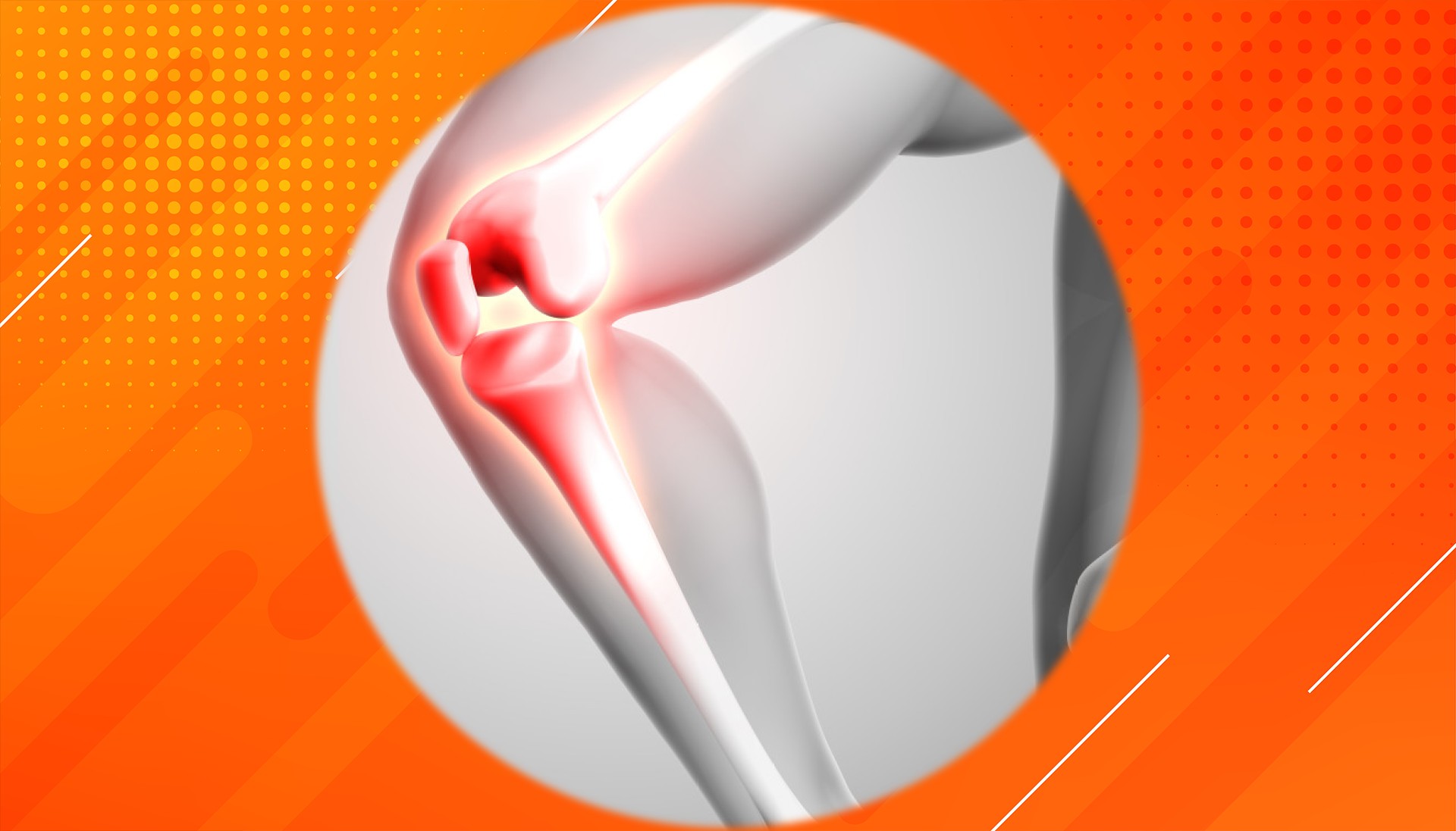
In osteoarthritis, the knee’s cartilage gradually wears down. In the early stages, the cartilage softens and thins, narrowing the space between the bones. As more cartilage is lost, bones start to rub against each other, leading to pain, swelling, and stiffness.
The body reacts by producing bony outgrowths called osteophytes (bone spurs) around the edges of the joint. While these can initially help stabilize the knee, over time they contribute to pain and limit movement.
All of these changes disrupt the knee’s normal movement and stability, creating a cycle where damage and attempted repairs lead to further joint problems. Early identification of these changes can help guide treatments that slow the progression of osteoarthritis.
How Biomechanics Affect Osteoarthritis
Several physical factors can increase the risk or worsen the progression of osteoarthritis. Poor knee alignment—such as being bow-legged or knock-kneed—causes body weight to concentrate on certain parts of the cartilage, speeding up its breakdown. When cartilage wears thin, its ability to cushion the joint diminishes, putting bones under greater direct stress.
This uneven loading changes the way the knee moves, straining nearby ligaments and muscles, and accelerating cartilage loss. Studies show that extra pressure on the inner or outer parts of the knee can hasten the onset and progress of osteoarthritis. The changes can be compounded in aging populations, where osteoarthritis becomes more common.
How the Knee Adapts to Stress
As osteoarthritis progresses, the knee attempts to adapt to these stresses. Bone spurs (osteophytes) form as a way to stabilize the joint, similar to adding extra supports to a weakening bridge. However, while initially helpful, these growths can eventually restrict movement and cause additional pain.
The loss of cartilage narrows the joint space, altering how weight travels through the knee. Ligaments may stretch or thicken in response to new patterns of movement and instability. While these adaptations temporarily support the knee, they often contribute to further dysfunction and pain as the disease advances.
These patterns of adaptation and degeneration are more common with age, particularly in women, and can occur in multiple joints—not just the knees.
What This Means for Treatment
By understanding the biomechanical factors that drive osteoarthritis, treatment can become more targeted and effective. Physical therapy is often recommended to strengthen the muscles around the knee, which supports joint stability and helps ensure forces are spread more evenly. Tools such as braces or specialized shoe inserts can re-align the knee during walking, reducing localized stress.
In more severe cases, surgical options may be necessary to realign the joint or replace damaged surfaces. Researchers are also exploring new treatments that act on a cellular level to protect or repair cartilage under mechanical stress. By addressing the underlying mechanics of knee osteoarthritis, doctors can create more personalized care plans—potentially delaying, or even avoiding, the need for joint replacement.
Looking Ahead: The Future of Knee Osteoarthritis Care
Studying how the knee responds to everyday forces provides invaluable insight into why osteoarthritis develops and how it worsens. As we continue to connect the dots between anatomy, biomechanics, and disease progression, new technologies—from advanced imaging to computer modeling—promise even deeper understanding.
These advances may unlock therapies that better protect knee structure, relieve pain, and restore mobility. As researchers and clinicians continue to explore this field, the future holds promise for improved quality of life for people affected by knee osteoarthritis.
References
- Magnusson, K., Kumm, J., Turkiewicz, A., & Englund, M. (2018). Early knee osteoarthritis or healthy ageing? Osteoarthritis and Cartilage, Abstract.